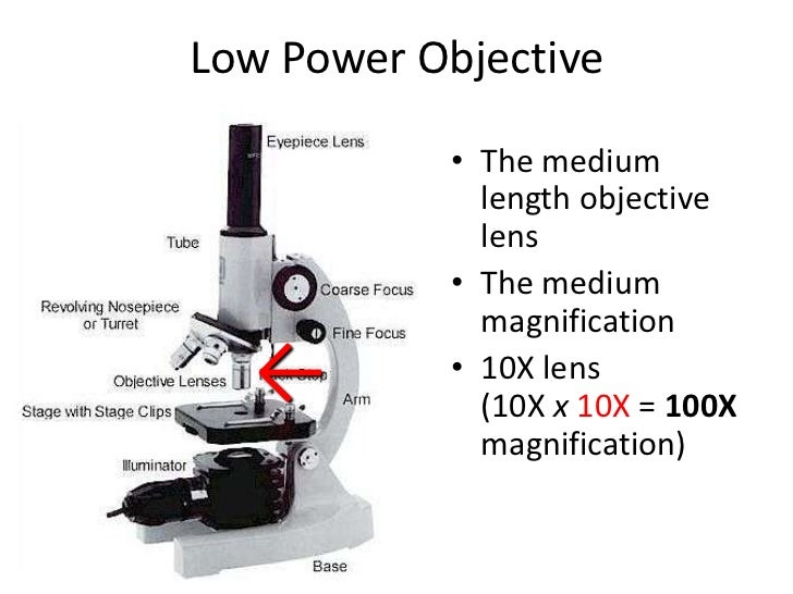
44 microscope parts labeled and functions
Parts of Binoculars and Their Functions Guide - TheOptics.org Binoculars help our eyes make sense of things that we would otherwise struggle to see by making them more prominent. This function is not powered by magic but by basic science. This is achieved by leveraging the optical laws known to man and the properties of light. However, for a binocular to work, it would need to be constructed right, and ... Microscope Types (with labeled diagrams) and Functions Simple microscope labeled diagram Simple microscope functions It is used in industrial applications like: Watchmakers to assemble watches Cloth industry to count the number of threads or fibers in a cloth Jewelers to examine the finer parts of jewelry Miniature artists to examine and build their work Also used to inspect finer details on products
What I Can Do A. Label the parts of the microscope below by putting ... Base: The bottom of the microscope, used for support. ... Revolving Nosepiece or Turret: This is the part of the microscope that holds two or more objective lenses and can be rotated to easily change power. Explanation: The Eyepiece Lens. ••• ... The Eyepiece Tube. ••• ... The Microscope Arm. ••• ... The Microscope Base ...
Microscope parts labeled and functions
Microscope, Microscope Parts, Labeled Diagram, and Functions 19.01.2022 · Microscope, Microscope Parts, Labeled Diagram, and Functions Published by Admin on January 19, 2022 January 19, 2022. What is Microscope? A microscope is a laboratory instrument used to examine objects that are too small to be seen by the naked eye. It is derived from Ancient Greek words and composed of mikrós, “small” and skopeîn,”to look” or … Microscope- Definition, Parts, Functions, Types, Diagram, Uses 21.02.2022 · History of Microscope. In the 1 st Century AD, the Romans invented the glass and used them to magnify objects.; In the early 14 th Century AD, eyeglasses were made by Italian spectacle makers.; In 1590, two Dutch spectacle makers, Hans, and Zacharias Jansen created the first microscope. It was a simple tube with 2 lenses system and had 9X magnification. UD Virtual Compound Microscope - University of Delaware ©University of Delaware. This work is licensed under a Creative Commons Attribution-NonCommercial-NoDerivs 2.5 License.Creative Commons Attribution-NonCommercial-NoDerivs 2.5 …
Microscope parts labeled and functions. Parts of Stereo Microscope (Dissecting microscope) – labeled … If you would like to learn optical components of a compound microscope, please visit Compound Microscope Parts – Labeled Diagram and their Functions, and this article. How to use a stereo (dissecting) microscope. Follow these steps to put your stereo microscopes in work: 1. Set your microscope on a tabletop or other flat sturdy surface where ... microscopewiki.com › compound-microscopeCompound Microscope – Diagram (Parts labelled), Principle and ... Feb 03, 2022 · This is the upper part of the microscope that houses the optical parts. 2. Arm . This part connects the head with the base and provides stability to the microscope. Arm is used to carry the microscope around. 3. Base . Base is on which the microscope rests and the base houses the illuminator that lights up the specimens. Optical Components Light Microscope Parts, Function & Uses - Study.com The following are the most common components found on light microscopes: Microscope parts Ocular lenses: Allow the viewer to look into the microscope, usually 10x magnification Revolving nosepiece:... rsscience.com › stereo-microscopeParts of Stereo Microscope (Dissecting microscope) – labeled ... If you would like to learn optical components of a compound microscope, please visit Compound Microscope Parts – Labeled Diagram and their Functions, and this article. How to use a stereo (dissecting) microscope. Follow these steps to put your stereo microscopes in work: 1. Set your microscope on a tabletop or other flat sturdy surface where ...
Label the parts of the microscope shown in the picture below using the ... Label the parts of the microscope shown in the picture below using the following terms: coarse adjustment knob, eyepiece (or ocular lens), fine adjustment knob, light condenser and iris diaphragm, objective lenses, stage ... All these parts are the important parts of microscope which has a specific function in the microscope. Without one of ... microscope parts and functions worksheet pdf - Blogger Microscope parts and functions worksheet answers. Label the parts Add to my workbooks 12 Download file pdf Embed in my website or blog Add to Google Classroom. Light Source Could be a mirror but most likely it is a bulb built into the base 2. Microscope parts and functions worksheet answer key. Never force the microscope parts to work. Compound Microscope Drawing With Parts and Functions The total magnification of image formed by the compound microscopes is calculated b this following formula; M = D/ fo * L/fe. Where, D = Least distance of distinct vision (25 cm) L = Length of the microscope tube. fo = Focal length of the objective lens. fe = Focal length of the eye-piece lens. researchtweet.com › microscope-parts-labeledMicroscope, Microscope Parts, Labeled Diagram, and Functions Jan 19, 2022 · Illuminator: Illuminator is the most important microscope parts and it serve as light source for a microscope during slide specimen visualization. It is a continuous source of light (110 volts) used in place of a mirror. The mirror of microscope is used to reflect light from the external light source up through the bottom of the stage.
microscope parts and functions worksheet pdf - Alverta Overton Microscope Parts and Functions. Lab work the student microscope name parts of environment light microscope review or fill almost the blanks wanganui high school. The lens at the top that you look through. Parts Of A Microscope With Functions And Labeled Diagram Microscope Parts Microscope Stain Techniques Inverted Microscope- Definition, Principle, Parts, Labeled … 10.04.2022 · Parts of a microscope with functions and labeled diagram Light Microscope- Definition, Principle, Types, Parts, Labeled Diagram, Magnification Amazing 27 Things Under The Microscope With Diagrams Light Microscope- Definition, Principle, Types, Parts, Labeled Diagram ... The functioning of the light microscope is based on its ability to focus a beam of light through a specimen, which is very small and transparent, to produce an image. The image is then passed through one or two lenses for magnification for viewing. The transparency of the specimen allows easy and quick penetration of light. Microscope slide - Wikipedia A microscope slide is a thin flat piece of glass, typically 75 by 26 mm (3 by 1 inches) and about 1 mm thick, used to hold objects for examination under a microscope.Typically the object is mounted (secured) on the slide, and then both are inserted together in the microscope for viewing. This arrangement allows several slide-mounted objects to be quickly inserted and …
microbenotes.com › inverted-microscopeInverted Microscope- Definition, Principle, Parts, Labeled ... Apr 10, 2022 · The working principle of the inverted microscope is basically the same as that of an upright light microscope. They use light rays to focus on a specimen, to form an image that can be viewed by the objective lenses. However, in the inverted microscope, the light source and the condenser are found on top of the stage pointing down to the stage.
thebiologynotes.com › microscopeMicroscope- Definition, Parts, Functions, Types, Diagram, Uses Feb 21, 2022 · Parts of a Microscope. A typical microscope contains the following parts; 1. Illuminator (Light Source) A microscopic illuminator is a light source. In some compound microscope, the mirror is used which reflect the light from an external source to the sample.
Parts of Stethoscope: A Comprehensive Overview 1-Headset. The headset is a part of a stethoscope, which is a combination of ear tips and ear tubes, and tension springs. These components combine together to fulfilling the purpose of diagnosis. It provides a comfortable alignment in the ears of a user and further used to provide maximum quality of sound through the headset.
Microscope: Types of Microscope, Parts, Uses, Diagram - Embibe There microscope anatomy includes three structural parts, i.e. head, base, and arm. Head - This is also known as the body; it carries the optical parts in the upper part of the microscope. Base - It acts as microscopes support. It also carries microscopic illuminators.
Electron Microscope Principle, Uses, Types and Images (Labeled … 02.02.2022 · Ans: A light microscope has a low resolving power (0.25µm to 0.3µm) while the electron microscope has a resolution power about 250 times higher than the light microscope at about 0.001µm. Similarly, a light microscope has a magnification of 500X to 1500x while the electron microscope has a much higher magnification of 100,000X to 300,000X.
Microscope Parts and Functions Microscope Parts and Functions With Labeled Diagram and Functions How does a Compound Microscope Work?. Before exploring microscope parts and functions, you should probably understand that the compound light microscope is more complicated than just a microscope with more than one lens.. First, the purpose of a microscope is to magnify a …
8 Parts of the Stethoscope, their Names, Functions, and Diagram The chest piece is also called as head of the stethoscope. It is a combination of diaphragm, bell, and stem. It is very much responsible for generating sound waves from vibrations in the body & transferring sound waves to the ear. To detect the vibrations chest piece is pressed on the back, chest, and stomach of the patient.
Epididymis Histology Slide and Identification Points with Labeled ... If you want to learn more about the anatomy of an epididymis, you may read another article from anatomy learner. Functions of epididymis ... Binocular Microscope Anatomy - Parts and Functions with a Labeled Diagram; Colon Histology Slide with Labeled Diagram; Cat Digestive System Anatomy with a Labeled Diagram;
Microscope Quiz: How Much You Know About Microscope Parts And Functions? It is used to support the microscope when carried. 2. Stage clips: A. Magnification ranges from 10x to 40x. B. Holds the slide in place. C. Moves the stage up and down for focusing. 3. Fine adjustment knob: A. Moves the stage slightly to sharpen the image. B. Holds the high and low power objectives. It can be rotated to change the magnification. C.


Post a Comment for "44 microscope parts labeled and functions"