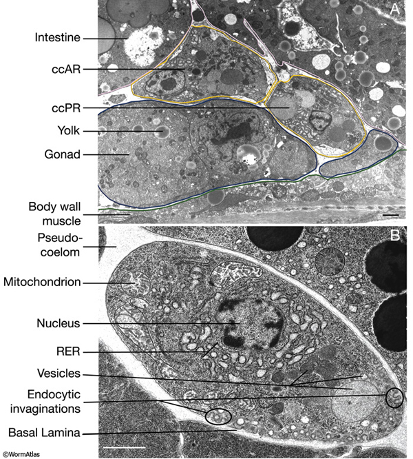
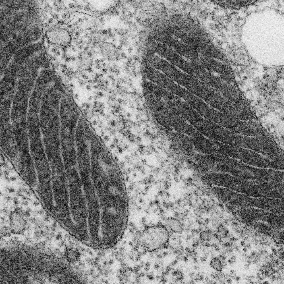
38 label the transmission electron micrograph of the mitochondrion.
Mitochondrial morphology and function: two for the price of one! This work represents a technical advance that allows the correlation of mitochondrial function and morphology with greater resolution and volume than has previously been feasible. LAY SUMMARY: Transmission electron microscopy (TEM) is a high-resolution technique used for the study of cells and their components, such as mitochondria. The influence of acetylsalicylic acid on oxylipin migration in ... Transmission electron microscopy suggests that osmiophilic material originates in mitochondria and is deposited inside the yeast cell wall, from which it is excreted into the environment, along capsule protuberances, or through capsule detachments.
Nelson Biology 12.pdf [30j71j2z320w] - doku.pub The orbital that is occupied by an electron is what determines the energy level of the electron. The farther away the electron is from the nucleus, the greater its energy. The balloon-like 2p orbitals contain electrons that are farther away from the nucleus than the electrons in the 2s orbital, and thus hold the electrons with a higher energy ...

Label the transmission electron micrograph of the mitochondrion.
Transmission electron microscopy techniques - University of Otago STEM (scanning transmission electron microscopy) enables the use of other of signals that cannot be spatially correlated in TEM, including secondary electrons, scattered beam electrons, characteristic X-rays, and electron energy loss—allowing very accurate elemental mapping and chemical analysis of samples. Single Particle Reconstruction Solved Label the transmission electron micrograph of the | Chegg.com Question: Label the transmission electron micrograph of the cell. 0 Nucleus rences Mitochondrion Heterochromatin Peroxisome Vesicle ULAR bumit Click and drag each label into the correct category to indicate whether it pertains to the cytoplasm or the plasma membrane. Microbiology Module 3 Flashcards - Quizlet The mitochondrion is the organelle involved in generating energy for the cell. It has a smooth continuous membrane surrounding its exterior. Inside, the inner membrane is folded into cristae. This inner membrane houses the proteins of the electron transport chain, involved in aerobic respiration. The fluid inside of the mitochondrion is called ...
Label the transmission electron micrograph of the mitochondrion.. Natural Sciences Grade 9 - mstworkbooks.co.za Remind your learners that a micrograph is a photograph taken through a microscope, and in this case, a transmission electron microscope. Left) A Diagram of a nucleus. ... Look at the micrograph of the mitochondrion in the image. ... Each organelle needs to be clearly labelled and with each label you need to add a description of the function of ... The Single Mitochondrion of Tachyzoites of Toxoplasma gondii Mitochondria from the thin serial sections of whole tachyzoites examined by host cell concentrate around the vacuolar membrane transmission electron microscopy further confirmed and are intensely labeled with rhodamine 123. One the observations on mitochondrial shape and num- possibility that could not be ruled out is that, for ber. Solved Label the transmission electron micrograph of the | Chegg.com Explanation - Mitochondrion is filamentous or globular in shape, occur in variable numbers from a few hundred to few thousands in different cells. It … View the full answer Transcribed image text: Label the transmission electron micrograph of the mitochondrion. Matrix granule Mitochondrion Outer membrane Cristae Inner membrane Matrix Reset Zoom The Transmission Electron Microscope | CCBER Transmission electron microscopes (TEM) are microscopes that use a particle beam of electrons to visualize specimens and generate a highly-magnified image. TEMs can magnify objects up to 2 million times. In order to get a better idea of just how small that is, think of how small a cell is. It is no wonder TEMs have become so valuable within the ...
Label This Transmission Electron Micrograph / Microscopy ... - Blogger Scanning Transmission Electron Micrograph Stem Vesicles Were Download Scientific Diagram from Labeling nuclear proteins with electron dense probes in living cells. 0 nucleus rences mitochondrion heterochromatin peroxisome vesicle ular bumit . The eluted conjugate is now ready for visualization by negative stain or cryo ... Electron Micrographs of Cell Organelles - Biology Discussion It is an electron micrograph of cell's largest and most important organelle - the mitochondria and is characterized by the following features (Fig. 7 & 8): (1) The name mitochondria was given by Benda (1898) and their ma n function was brought to light by Kingsbury (1912). Native Immunogold Labeling of Cell Surface Proteins and Viral ... Introduction. Cryo-electron microscopy (cryo-EM) technologies and techniques were established in order to retain specimen hydration during direct-image and diffraction data acquisition with the transmission electron microscope (TEM) (Taylor and Glaeser 1974).Much of the early work in the late 1970s and early 1980s focused on improving cryo-fixation approaches (Dubochet et al. 1988; Lepault et ... Looking at the Structure of Cells in the Microscope Determining the detailed structure of the membranes and organelles in cells requires the higher resolution attainable in a transmission electron microscope. Specific macromolecules can be localized with colloidal gold linked to antibodies. Three-dimensional views of the surfaces of cells and tissues are obtained by scanning electron microscopy.
Brit A Z | PDF | Nature - Scribd aardvark aardvarks aardvark's aardwolf ab abaca aback abacus abacuses abaft abalone abalones abalone's abandon abandoned abandonee. abandoner abandoning abandonment abandons abase abased abasement abasements abases abash abashed abashes abashing abashment abasing abate abated abatement abatements abates abating abattoir abbacy … Light microscopy of mitochondria at the nanoscale - PMC The unique membrane architecture of mitochondria was discovered in the 1950s by the use of transmission electron microscopy, at the time a new enabling technology for cell biology ( 6; 7) ( Fig. 1 ). › 37006818 › Junqueiras_BasicJunqueira's Basic Histology Text and Atlas, 14th Edition Enter the email address you signed up with and we'll email you a reset link. Label the transmission electron micrograph of the nucleus. Label the transmission electron micrograph of the cell. 0 Nucleus rences Mitochondrion Heterochromatin Peroxisome Vesicle ULAR bumit Click and drag each label into the correct category to indicate whether it pertains to the cytoplasm or the plasma...
› pmc › articlesCryo-electron microscopy: A primer for the non-microscopist Over the past decade, the phrase “cryo-electron microscopy”, often abbreviated as “cryo-EM”, has evolved to encompass a broad range of experimental methods. At the core, each of these is based upon the principle of imaging radiation-sensitive specimens in a transmission electron microscope under cryogenic conditions.
Biology Exam Review Flashcards | Quizlet The table below contains an electron micrograph of each of the commonly occurring organelles, with a drawing of the structure. Brief notes on recognition features and the function of each organelle are included. ... Mitochondrion. A double membrane surrounds mitochondria, with the inner of these membranes invaginated to form structures called ...
Visualizing Mitochondrial Form and Function within the Cell While there are several methods for measuring cellular mitochondrial content [2, 13-17], imaging is the only way to directly assess the volume of a cell that is occupied by mitochondria. For light microscopy, there are three general approaches to visualizing mitochondrial structures: 1) labeling mitochondria in live cells with fluorescent ...
doku.pub › documents › nelson-biology-12pdf-30j71j2z320wNelson Biology 12.pdf [30j71j2z320w] - doku.pub The orbital that is occupied by an electron is what determines the energy level of the electron. The farther away the electron is from the nucleus, the greater its energy. The balloon-like 2p orbitals contain electrons that are farther away from the nucleus than the electrons in the 2s orbital, and thus hold the electrons with a higher energy ...
› 44090147 › CambridgeCambridge International AS and A Level Biology Coursebook ... Enter the email address you signed up with and we'll email you a reset link.
Analysis of Yeast Mitochondria by Electron Microscopy We provide here detailed protocols for the analysis of yeast mitochondria by transmission electron microscopy: (1) chemical fixation and Epon embedding of yeast cells and isolated mitochondria, and (2) cryosectioning and immunolabeling of yeast cells and isolated mitochondria according to the Tokuyasu method. less
Transmission Electron Microscopy for Analysis of Mitochondria in Mouse ... Transmission electron microscopy (TEM) is a powerful technique for ultrastructural studies ( Watson, 1958 ). TEM has been very useful in studying mitochondrial structure in skeletal muscle in both physiological and pathological conditions ( Picard et al., 2013 ).
Localization of A44 within mitochondria. Shown is transmission electron ... Download scientific diagram | Localization of A44 within mitochondria. Shown is transmission electron microscopy of sections of host cells either expressing A44-GFP (A) or incubated with exogenous ...
PDF Identifying Organelles from an Electron Micrograph ELECTRON MICROGRAPH OF MITOCHONDRION Courtesy of Electron Microscopy Unit University of Lancaster endoplasmic cristae reticulum fluid matrix outer membrane ER, cristae, fluid matrix, ribosome, outer membrane Courtesy of Dr. Julian Thorpe - EM & FACS Lab, Biological Sciences University Of Sussex
Solved Label the transmission electron micrograph based on - Chegg Expert Answer nucleus is the house of the genetic material which contains all the h … View the full answer Transcribed image text: Label the transmission electron micrograph based on the hints provided Mitochondrion Heterochromatin Plasma cell Nucleus Rough endoplasmic reticulum Nucleolus Previous question Next question
Electron Micrographs** Electron Micrographs**. Below is a collection of electron micrographs with labelled subcellular structures that you should be able to identify. Also, be sure to observe any electron micrographs which are made available in the laboratory by the instructor. You should concentrate on the similarities in form that permit identification of the ...
Frontiers | Mitochondrial Morphology and Mitophagy in Heart Diseases ... Transmission electron microscopy (TEM) has long been an important technique, capable of high degree resolution and visualization of subcellular structures and organization. Over the last 20 years, TEM has gained popularity in the cardiovascular field to visualize changes at the nanometer scale in cardiac ultrastructure during cardiovascular development, aging, and a broad range of pathologies.
Draw the structure of a mitochondrion as seen in an electron micrograph ... Last Updated 13 Jul 2020 Draw the structure of a mitochondrion as seen in an electron micrograph 411 6)a) Draw the structure of a mitochondrion as seen in an electron micrograph. [5] B) Describe the central role of acetyl (ethanoyl) CoA in carbohydrate & fat metabolism. [5] Acetyl CoA is formed in both carbohydrate and fat metabolism.
00 (1).pdf - Practice 1.2 [48 marks] 1a./1 mark] Identify ... - Course Hero View 00 (1).pdf from CHEM-UA MISC at New York University. Practice 1.2 [48 marks] 1a./1 mark] Identify which electron micrograph shows a mitochondrion, providing one observation to support your
AICE Biology Chapter 1: Plant Cell Electron Micrograph Labeling Start studying AICE Biology Chapter 1: Plant Cell Electron Micrograph Labeling. Learn vocabulary, terms, and more with flashcards, games, and other study tools.
Cambridge International AS and A Level Biology Coursebook … Enter the email address you signed up with and we'll email you a reset link.
Mitochondria under the microscope - Science Learning Hub Microscopes have been crucial for our understanding of mitochondrial structure and function. Mitochondria are visible under the light microscope although little detail can be seen. Transmission electron microscopy (left) shows the complex internal membrane structure of mitochondria, and electron tomography (right) gives a three-dimensional view.
medicalxpress.com › news › 2022-06-uncover-host-cellResearchers uncover a novel host cell pathway hijacked during ... Jun 14, 2022 · An international team of scientists, led by the University of Bristol, has been investigating how the SARS-CoV-2 virus, the coronavirus responsible for the COVID-19 pandemic, manipulates host ...
Labeling the Cell Flashcards - Quizlet Label the transmission electron micrograph of the mitochondrion. Label the transmission electron micrograph of the nucleus. membrane bound organelles golgi apparatus, mitochondrion, lysosome, peroxisome, rough endoplasmic reticulum nonmembrane bound organelles ribosomes, centrosome, proteasomes cytoskeleton includes
Bio101 - Ch 6 HW Flashcards - Quizlet Drag the labels on the left onto the diagram of the animal cell to correctly identify the function performed by each cellular structure. a. smooth ER- synthesizes lipids b. nucleolus- assembles ribosomes c. defines cell shape d. rough ER- produces secretory proteins e. Golgi apparatus- modifies and sorts proteins f. digests proteins
quizlet.com › ca › 259010575Biology Exam Review Flashcards - Quizlet A photo taken down a microscope is called a micrograph. There are many micrographs in this book, including electron micrographs taken using an electron microscope. When we draw a specimen, we can make the drawing larger or smaller, so the magnification of the drawing isn't necessarily the same as the magnification of the microscope.
Schaums Outline Of Genetics [PDF] [1vrels341bno] - vdoc.pub An organelle present in animal cells but missing from plant cells is (c) a vacuole ( d ) a mitochondrion (e) more than one of the above (a) a nucleolus (6) a centriole 2. How many spermatids are normally produced by 50 primary spermatocytes? I d ) 200 (e) 400 (a) 25 (6) 50 (c) 100 3. Humans normally have 46 chromosomes in skin cells.
Junqueira's Basic Histology Text and Atlas, 14th Edition Enter the email address you signed up with and we'll email you a reset link.
› natural-sciences › gr9Natural Sciences Grade 9 - Grade 7-9 Workbooks What differences can you see between this mitochondrion and the diagram shown previously? In the diagram, it was very clear that the inner membrane folds, whereas in the micrograph it is not as easy to see this. This is because of the way that the cell was sectioned (cut) before it was viewed on the transmission electron microscope.
Microbiology Module 3 Flashcards - Quizlet The mitochondrion is the organelle involved in generating energy for the cell. It has a smooth continuous membrane surrounding its exterior. Inside, the inner membrane is folded into cristae. This inner membrane houses the proteins of the electron transport chain, involved in aerobic respiration. The fluid inside of the mitochondrion is called ...
Solved Label the transmission electron micrograph of the | Chegg.com Question: Label the transmission electron micrograph of the cell. 0 Nucleus rences Mitochondrion Heterochromatin Peroxisome Vesicle ULAR bumit Click and drag each label into the correct category to indicate whether it pertains to the cytoplasm or the plasma membrane.
Transmission electron microscopy techniques - University of Otago STEM (scanning transmission electron microscopy) enables the use of other of signals that cannot be spatially correlated in TEM, including secondary electrons, scattered beam electrons, characteristic X-rays, and electron energy loss—allowing very accurate elemental mapping and chemical analysis of samples. Single Particle Reconstruction












Post a Comment for "38 label the transmission electron micrograph of the mitochondrion."