40 correctly label the following internal anatomy of the heart
Chapter 20-Cardiovascular System Flashcards - Quizlet Memorize flashcards and build a practice test to quiz yourself before your exam. Start studying the Chapter 20-Cardiovascular System flashcards containing study terms like Correctly label the following internal anatomy of the heart., Correctly label the following internal anatomy of the heart. b, Place the labels in order denoting the flow of oxygenated blood through the heart beginning with ... [Solved] Check my work Correctly label the followiting Internal anatomy ... E correcty label the following Internal anatomy of the heart Left atrium Interventricular septum Papillary muscles Right ventricle opening of superior Left ventricle vena cava Fossa ovalis right atrium Right atrium Pectinate tricuspid valve muscles right ventricle rabeculae camae papillary muscle O < Prev 5 of 25 Next > Download Attachments ...
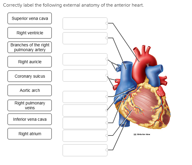
Correctly label the following internal anatomy of the heart. Right ... Saved Correctly label the following external anatomy of the anterior heart. 18 Apex of heart 0.37 points Ascending aorta Skipped Ligamentum arteriosum Left pulmonary veins References Pulmonary trunk Anterior interventricular sulous Left auricle Left...

Correctly label the following internal anatomy of the heart
Sheep Heart Dissection Lab for High School Science | HST Dissection: Internal Anatomy. 1. Insert your dissecting scissors or scalpel. into the superior vena cava and make an incision down through the wall of the right atrium and ventricle, as shown by the dotted line in the external heart picture. Pull the two sides apart and look for three flaps of the membrane. Solved Correctly label the following internal anatomy of the Question: Correctly label the following internal anatomy of the heart. Left pulmonary veins Left ventricle Myocardium Endocardium Pulmonary trunk ...1 answer · Top answer: Starting from Top right to bottom.... Lesson | The Heart - External Structure | Encounter Edu Veins appear blue through the skin because of refraction but they are actually dark red. Students label the diagram of the external structure of the heart as they explore the heart and read the information (see Student Sheet found in the Lesson resources section). Students answer the questions in the scene and discuss their ideas with partners.
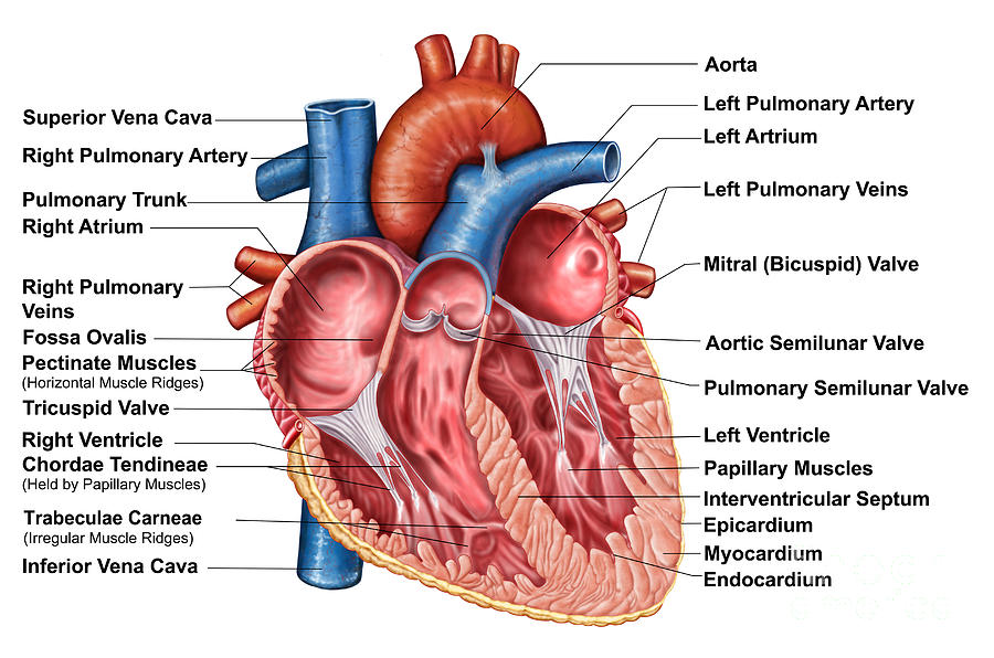
Correctly label the following internal anatomy of the heart. Layers of the heart: Epicardium, myocardium, endocardium - Kenhub The myocardium is functionally the main constituent of the heart and the thickest layer of all three heart layers. It is a muscle layer that enables heart contractions. Histologically, the myocardium is comprised of cardiomyocytes.Cardiomyocytes have a single nucleus in the center of the cell, which helps to distinguish them from skeletal muscle cells that have multiple nuclei dispersed in the ... Label the heart - Science Learning Hub In this interactive, you can label parts of the human heart. Drag and drop the text labels onto the boxes next to the heart diagram. If you want to redo an answer, click on the box and the answer will go back to the top so you can move it to another box. If you want to check your answers, use the Reset Incorrect button. Solved Correctly label the following parts of the internal - Chegg Correctly label the following parts of the internal anatomy of the heart. Right pulmonary veins Aorta Left pulmonary veins Pulmonary semilunar Left ventricle Right ventricle Bicuspid valve Tricuspid valve Right pulmonary artery Septum Pulmonary trunk Right atrium Left pulmonary artery Inferior vena cava Left atrium Superior vena cava Reset Zoom ... Quiz 4 - Quiz4 1. Award: 0 out of 1.00 point Classify the following ... Award: 1 out of 1.00 point Correctly label the following parts of the internal anatomy of the heart. References Labeling Section: 05.03 3. Award: 1 out of 1.00 point Using the image as a guide, sequence the following descriptions of the flow of blood through the human heart.
Heart Labeling Quiz: How Much You Know About Heart Labeling? Here is a Heart labeling quiz for you. The human heart is a vital organ for every human. The more healthy your heart is, the longer the chances you have of surviving, so you better take care of it. Take the following quiz to know how much you know about your heart. 1. Solved Correctly label the following internal anatomy of the - Chegg Anatomy and Physiology questions and answers. Correctly label the following internal anatomy of the heart. Right atrium Pectinate muscles Trabeculae carnae Left ventricle Fossa ovalis Right ventricle Interventricular seplum Left atrium Papiliary muscles Reset Zoom. Question: Correctly label the following internal anatomy of the heart. The Anatomy of the Heart, Its Structures, and Functions The heart is the organ that helps supply blood and oxygen to all parts of the body. It is divided by a partition (or septum) into two halves. The halves are, in turn, divided into four chambers. The heart is situated within the chest cavity and surrounded by a fluid-filled sac called the pericardium. This amazing muscle produces electrical ... Label Parts of the Human Ear - University of Dayton Label Parts of the Human Ear. Select One Auditory Canal Cochlea Cochlear Nerve Eustachian Tube Incus Malleus Oval Window Pinna Round Window Semicircular Canals Stapes Tympanic Membrane Vestibular Nerve. Select One Auditory Canal Cochlea Cochlear Nerve Eustachian Tube Incus Malleus Oval Window Pinna Round Window Semicircular Canals Stapes ...
Anatomy & Physiology: The Unity of Form and Function - Quizlet Correctly label the following external anatomy of the posterior heart. Correctly label the following internal anatomy of the heart. Correctly sequence the pathway of blood flow through the heart, beginning with the venae cavae. Correctly label the following internal anatomy of the heart. Fossa ... Correctly label the following parts of the internal anatomy of the heart. Place your cursor over the boxes for more information papillary muscles bicuspid valve right atrium septum pulmonary semilunar valve eft atrium chordae tendineae pulmonary... Heart Dissection Walk Through - The Biology Corner Don't be shy with the heart, use your fingers to feel your way through the dissection. 1. Step One: Orientation. When you first remove your heart from the bag, you will see a lot of fatty tissue surrounding it. It is usually a waste of time to try to remove this tissue. Grab some colored pencils to help you identify and mark the vessels you find. Heart Anatomy: Labeled Diagram, Structures, Blood Flow ... - EZmed There are 4 chambers, labeled 1-4 on the diagram below. To help simplify things, we can convert the heart into a square. We will then divide that square into 4 different boxes which will represent the 4 chambers of the heart. The boxes are numbered to correlate with the labeled chambers on the cartoon diagram.
Heart Anatomy: chambers, valves and vessels : Anatomy & Physiology
11 A&p ideas | anatomy and physiology, physiology, human anatomy and ... Feb 13, 2019 - Explore Imissthe Oldme's board "a&p" on Pinterest. See more ideas about anatomy and physiology, physiology, human anatomy and physiology.

Electrocardiogram (ECG, EKG) Causes, Symptoms, Treatment - Basic ...
Answered: Correctly label the following veins of… | bartleby Correctly label the following veins of the thorax. Hemiazygos v. Internal jugular v. Subclavian v. Posterior intercostal veins SubclaviarM Brachiocephalic V. Supreme intercostal v. Azygos v. Internal jugular v.
31 Label This Anterior View Of The Human Heart - Labels Database 2020
Structure of the Heart | SEER Training Structure of the Heart. The human heart is a four-chambered muscular organ, shaped and sized roughly like a man's closed fist with two-thirds of the mass to the left of midline. The heart is enclosed in a pericardial sac that is lined with the parietal layers of a serous membrane. The visceral layer of the serous membrane forms the epicardium.
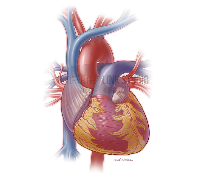
Heart Anatomy External
25.1 Internal and External Anatomy of the Kidney The left kidney is located at about the T12 to L3 vertebrae, whereas the right is lower due to slight displacement by the liver. Upper portions of the kidneys are somewhat protected by the eleventh and twelfth ribs ( Figure 25.1.1 ). Each kidney weighs about 125-175 g in males and 115-155 g in females. They are about 11-14 cm in length, 6 ...

36 Label Heart Parts - Labels 2021
Heart Blood Flow | Simple Anatomy Diagram, Cardiac Circulation ... - EZmed One of the first things you will notice if you look at the 12 steps is the pattern between the right and left side of the heart is similar. Step 1 and 6 involve a blood vessel, which makes sense as this is how blood enters and exits that side of the heart. Steps 2-5 involve a chamber, valve, chamber, and valve.

Februari 2011
The Heart - Science Quiz - GeoGuessr The Heart - Science Quiz: Day after day, your heart beats about 100,000 times, pumping 2,000 gallons of blood through 60,000 miles of blood vessels. If one of your organs is working that hard, it makes sense to learn about how it functions! This science quiz game will help you identify the parts of the human heart with ease. Blood comes in through veins and exists via arteries—to control the ...
Anatomy Of Heart Interior, Frontal Digital Art by Stocktrek Images
Diagrams, quizzes and worksheets of the heart - Kenhub Worksheet showing unlabelled heart diagrams. Using our unlabeled heart diagrams, you can challenge yourself to identify the individual parts of the heart as indicated by the arrows and fill-in-the-blank spaces. This exercise will help you to identify your weak spots, so you'll know which heart structures you need to spend more time studying ...
/heart_interior-570555cf3df78c7d9e908901.jpg)
Heart Wall: Epicardium, Myocardium, and Endocardium
Chapter 11 Cardiovascular System Answers Start studying the Chapter 20-Cardiovascular System flashcards containing study terms like Correctly label the following internal anatomy of the heart., Correctly label the following internal anatomy of the heart. b, Place the labels in order denoting the flow of oxygenated blood through the heart …

Heart Anatomy External
Cardiovascular System Heart Chapter 20 - hex.arista.com following internal anatomy of the heart., Correctly label the following internal anatomy of the heart. b, Place the labels in order denoting the flow of oxygenated blood through the heart beginning with Simplified relationship of the serous pericardium to the heart. A B. 326 CHAPTER 11 The CardiovascularSystem The Heart Ensures Continual, 24/7 ...
Post a Comment for "40 correctly label the following internal anatomy of the heart"